


, trigger signaling that strengthens proteostasis and stress resistance rather than causing damage. ...&media=http://cdn//csuxjmfbwmkxiegfpljm.supabase.co/storage/v1/object/public/blog-images/organization-27528/1789454429077_What-Role-Does-Mitohormesis-Play-in-Aging-overview-diagram.jpeg)


Mitohormesis is the adaptive response in which low doses of mitochondrial stress, mostly mild bursts of reactive oxygen species (mtROS), trigger signaling that strengthens proteostasis and stress resistance rather than causing damage. The same stress at high, sustained doses does the opposite: it degrades mitochondrial function and accelerates decline. For researchers and clinicians, this dose dependency is the mechanistic thread connecting exercise, dietary restriction, and select drugs to healthspan extension, and it sets up everything that follows: mechanisms, evidence across species, interventions, biomarkers, and where the risks actually sit.
TL;DR:
- Moderate mitochondrial stress from exercise or fasting activates protective pathways, but high or chronic stress flips the switch to damage and decline.
- Biomarkers like GDF15, FGF21, and mitochondrial peptides help track adaptive responses, yet their levels must be interpreted within specific timing and context.
- Nutritional supplements supporting redox balance, such as Nrf2 activators, can aid resilience but should complement, not replace, lifestyle interventions like exercise and fasting.
- Chronic overexposure to mitochondrial stress signals may promote inflammation and tumor survival, emphasizing the importance of dose and recovery timing.
- Interventions should focus on dose, timing, and individual variability, avoiding high-dose antioxidants around stress events to preserve the mitohormetic signaling.
Table of Contents
- What Is Mitohormesis at the Molecular Level?
- What Evidence Supports Mitohormesis Across Species?
- How Do Exercise, Fasting, and Drugs Trigger Mitohormesis?
- What Biomarkers Detect Mitohormesis in the Body?
- Where Does Mitohormesis Help, and Where Could It Hurt?
- Who Is Behind This Research Framing?
- How Was Mitohormesis Discovered?
- How Does Mitohormesis Relate to General Hormesis?
- What Role Does Mitohormesis Play in Aging?
- How Does Mitohormesis Affect Metabolic Health?
- Practical Takeaways for Research or Clinical Use
- Where Supplements Fit Alongside Mitochondrial-Support Habits
- Sources
- FAQ
What Is Mitohormesis at the Molecular Level?
Hormesis follows a biphasic, U-shaped dose-response curve. A small stressor, like a brief spike in mtROS during exercise, activates protective gene programs. A large or chronic stressor overwhelms those same defenses and produces oxidative damage, inflammation, and cell death. The direction of the outcome depends entirely on magnitude, duration, and cellular compartment, not on ROS presence alone.
Two coordinated stress programs translate that signal into durable adaptation. The mitochondrial unfolded protein response (UPRmt) senses misfolded matrix proteins and activates ATF-family transcription factors to boost chaperone and protease expression. The integrated stress response (ISR), regulated partly through the OMA1/DELE1 axis, slows general translation while up regulating stress-resistance transcripts. Both pathways converge on improved protein quality control.
Downstream, mitophagy clears damaged organelles, while fission and fusion dynamics redistribute damaged components for either repair or removal. Mitochondrial-derived peptides such as MOTS-c and humanin act as additional effectors, signaling to the nucleus and to distant tissues.
Researchers commonly track:
- UPRmt-associated chaperone and protease transcripts
- ISR markers, including ATF4 and downstream targets
- Mitophagy flux assays and fission/fusion morphology
- Circulating MOTS-c and humanin levels
What Evidence Supports Mitohormesis Across Species?
The strongest early support came from invertebrate models. Mild ROS elevation in C. elegans extends lifespan when mitochondrial electron transport is partially inhibited, an effect reversed by antioxidant co-treatment, a pattern replicated in Drosophila studies of dietary and genetic mitochondrial perturbation. Rodent work extended the concept: conplastic mice with mismatched mitochondrial and nuclear genomes show altered stress signaling, and exercise reliably induces UPRmt activation in mammalian skeletal muscle.
What the numbers show: intervention studies find that non-specific, high-dose antioxidant supplementation frequently blunts the benefits of exercise, and antioxidant enzyme overexpression does not reliably extend lifespan in the same animal models where mild ROS elevation succeeds. That asymmetry is one of the more counterintuitive findings in the field.
Human data remain more circumstantial. Urolithin A is being studied in clinical trials for its mitophagy-stimulating effects, and metformin’s mild inhibition of electron transport is associated with systemic metabolic signals consistent with a mitohormetic mechanism. Translation from worm to human is complicated by differences in measurement timing, tissue accessibility, and baseline metabolic rate, so causal claims in humans should stay cautious even where the biology is plausible.
How Do Exercise, Fasting, and Drugs Trigger Mitohormesis?
Real-world interventions work by nudging mitochondria into that narrow beneficial stress zone, and the variables that matter are dose, timing, and which tissue is exposed.
- Exercise produces a Goldilocks window. Moderate-to-high intensity training reliably activates UPRmt and mitokine release in skeletal muscle, but chronic overtraining without recovery pushes past the adaptive range into damage.
- Dietary restriction and intermittent fasting create transient mtROS pulses tied to metabolic rewiring, shifting cells toward fatty-acid oxidation and away from constant substrate availability.
- Pharmaceutical and nutraceutical triggers act through distinct mechanisms: metformin causes mild electron transport chain inhibition, urolithin A stimulates mitophagy directly, and Nrf2-activating phytochemicals, including many polyphenols, amplify endogenous antioxidant gene expression rather than scavenging ROS outright.
Pro Tip: If you’re designing or advising on a mitohormetic protocol, separate high-dose antioxidant supplementation from the stress-inducing intervention by several hours. Taking large doses of vitamin C or E immediately around a workout is one of the most common ways people accidentally cancel the adaptive signal they were trying to create.
Context determines outcome here more than any single ingredient does, a point covered in more depth in this discussion of redox signaling for clinicians and researchers.
What Biomarkers Detect Mitohormesis in the Body?
Two circulating mitokines dominate current research: GDF15 and FGF21. Both increase after mitochondrial stress and have different temporal profiles, with GDF15 more closely tied to acute stress signaling and systemic metabolic effects, and FGF21 more associated with sustained metabolic adaptation. Neither is specific to mitohormesis alone, so elevated levels need context from the rest of a study design.
Useful readouts for research or translational work include:
- MOTS-c and other mitochondrial-derived peptides, detectable via targeted assays but sensitive to sample handling
- UPRmt gene expression signatures in accessible tissue, when biopsy is feasible
- F2-isoprostanes as an oxidative stress readout, interpreted alongside mitokine data rather than alone
- Time-series sampling, since an acute rise that returns toward baseline within hours to days looks more like adaptive signaling than chronic elevation does
Where Does Mitohormesis Help, and Where Could It Hurt?
The same signaling pathways that support resilience can, if activated chronically, support processes researchers would rather suppress.
Plausible benefit sits in metabolic disease management, skeletal muscle resilience during aging, ischemic preconditioning, and general age-related functional decline. The concern runs in the opposite direction: chronic activation of mitohormetic pathways may support tumor cell survival and, in some contexts, metastatic potential, since cancer cells can hijack the same stress-adaptation machinery that protects healthy tissue.
- Non-specific antioxidant supplementation often fails or backfires because ROS effects are compartment- and time-dependent, and blanket scavenging removes the signal along with the damage.
- Biomarker-guided, temporally optimized interventions are a more defensible research direction than fixed-dose protocols applied uniformly across patients.
Who Is Behind This Research Framing?
This explainer is written by cristopher for Superiorformulas, drawing on peer-reviewed reviews of mitochondrial stress signaling; see the author bio for full credentials. For deeper technical reading, Superiorformulas maintains resources on cellular antioxidants and longevity and mitochondrial contributions to aging. Superiorformulas manufactures in GMP-certified facilities with third-party testing, a baseline transparency standard relevant to anyone evaluating mitochondrial-support products.
How Was Mitohormesis Discovered?
The term “mitohormesis” formalized an observation that had been accumulating for decades in redox biology: mild oxidative stress sometimes improves organismal outcomes rather than harming them. This ran against the dominant free radical theory of aging, which framed ROS purely as damaging byproducts to be neutralized.
Early evidence came from caloric restriction studies in the 1990s and 2000s, where lifespan extension in yeast, worms, and rodents correlated with modest increases in mitochondrial respiration and ROS production rather than their suppression. Researchers working with C. elegans found that partially inhibiting electron transport chain components, which would be expected to reduce ROS and extend lifespan under the old model, sometimes shortened it instead, while other genetic manipulations that mildly increased ROS extended it.
The conceptual breakthrough came when investigators demonstrated that antioxidant co-treatment could reverse the lifespan benefits of mild mitochondrial stress in these models. That single finding forced a rethink: ROS were acting as signaling molecules, not just damage agents, a distinction now central to how mitochondrial stress signaling is understood across the field.
Since then, the concept has expanded from a niche observation in invertebrate aging research into a framework applied to exercise physiology, pharmacology, and nutraceutical development. The shift has been gradual rather than sudden, and mainstream clinical practice still lags behind the mechanistic literature by a fair margin.
How Does Mitohormesis Relate to General Hormesis?
Mitohormesis is a specific case of the broader hormesis principle, applied to one organelle rather than to the cell or organism as a whole. General hormesis describes any biphasic dose-response relationship, where low doses of a stressor (radiation, heat, toxins, exercise) produce beneficial adaptive effects while high doses cause harm. Mitohormesis narrows that lens to mitochondrial stressors specifically, mostly mtROS, and to the organelle-specific signaling programs, UPRmt, ISR, and mitophagy, that mediate the adaptive response.
The relationship matters because mitochondria sit at a unique crossroads: they generate the bulk of cellular ROS as a byproduct of oxidative phosphorylation, they contain their own genome, and they communicate with the nucleus and with distant tissues through mitokines. That combination gives mitochondrial stress a broader signaling reach than, say, a localized heat-shock response confined to cytosolic proteostasis. A mitohormetic signal can influence gene expression in the nucleus, alter systemic metabolism through circulating peptides, and affect neighboring cells, all from a single organelle-level perturbation.
Practically, this means mitohormesis inherits the core rules of general hormesis, dose matters more than presence or absence of the stressor, timing matters, and chronic overexposure flips the outcome, while adding organelle-specific complexity. A researcher studying skin hormesis to UV exposure and a researcher studying skeletal muscle mitohormesis from exercise are working with the same underlying dose-response logic, but the mitochondrial case carries systemic reach that most other hormetic responses do not.
What Role Does Mitohormesis Play in Aging?
Aging research has moved away from a simple “ROS causes aging” model toward a framework where mitochondrial stress signaling actively shapes the pace of decline, for better or worse depending on context. Mitohormesis offers a mechanistic bridge between several established longevity interventions and their downstream effects on cellular resilience.
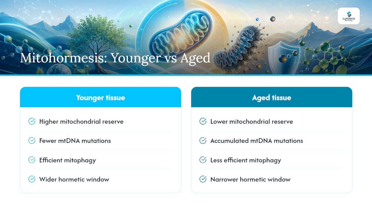
Caloric restriction, one of the most reproducible lifespan-extension interventions across model organisms, appears to work partly through transient mtROS elevation that triggers protective UPRmt and ISR activation rather than through simple metabolic slowdown. Exercise-induced longevity benefits follow a similar logic: repeated mild mitochondrial stress from training builds a more resilient proteostasis network over time, evident in the mitokine and gene-expression signatures researchers track after training bouts. The aging-relevant piece is that mitochondrial function declines with age on its own, reduced biogenesis, accumulated mitochondrial DNA mutations, and less efficient mitophagy all show up in older tissue. This creates a paradox worth sitting with: aged tissue may have a narrower hormetic window, meaning a stress dose that was beneficial at 30 could tip toward harmful at 70, because baseline mitochondrial reserve is lower and quality-control mechanisms are less responsive. That narrowing window is likely one reason fixed-intensity exercise or fasting protocols don’t produce identical benefits across age groups, and it argues for age-adjusted dosing in both research design and clinical recommendations rather than one-size-fits-all protocols.
How Does Mitohormesis Affect Metabolic Health?
Metabolic disease pathways intersect with mitohormesis at nearly every level, from insulin sensitivity to hepatic fat metabolism. The mitokines GDF15 and FGF21, both mitohormetic signaling outputs, have direct effects on systemic energy balance: FGF21 promotes fatty acid oxidation and improves insulin sensitivity in several mammalian studies, while GDF15 influences appetite regulation and has been studied as a marker of metabolic stress in conditions ranging from obesity to mitochondrial disease.
Metformin’s role here is instructive. Its mild inhibition of complex I in the electron transport chain induces a low-level mitochondrial stress response that appears to contribute to its glucose-lowering effects, independent of its more commonly cited action on hepatic glucose production. That mechanism places metformin squarely within the mitohormesis framework rather than treating it as a metabolic drug with an unrelated mode of action.
Skeletal muscle, as the body’s largest metabolically active tissue, is where exercise-induced mitohormesis likely has its biggest metabolic payoff. Repeated bouts of moderate exercise stress trigger mitochondrial biogenesis and improved oxidative capacity, which over time improves whole-body glucose disposal and lipid handling. This is a large part of why exercise remains one of the most effective interventions for insulin resistance and type 2 diabetes risk, even independent of weight loss.
The disease-pathway risk sits on the other side of the same coin: chronic, unresolved mitochondrial stress, the kind seen in prolonged overnutrition or sedentary metabolic dysfunction, tends to push mitokine signaling toward a maladaptive, inflammatory state rather than a protective one. Distinguishing acute, adaptive mitokine elevation from chronic, pathological elevation is one of the harder open problems in translating this biology into metabolic disease treatment.
Practical Takeaways for Research or Clinical Use
Three priorities stand out for anyone applying this framework. First, design studies or interventions around dose and timing, not just presence of a stressor, since the same intervention can help or harm depending on intensity and recovery windows. Second, use mitokine and UPRmt biomarkers to guide protocols rather than relying on symptom response alone. Third, keep high-dose antioxidants away from the stress window entirely.
For patient conversations, the simplest honest framing is that mild stress can build resilience, but more is not automatically better, and biomarkers help find the right dose for that individual.
— cristopher
Where Supplements Fit Alongside Mitochondrial-Support Habits
Nutritional support can complement, but never replace, the lifestyle interventions that actually generate a mitohormetic signal. Certain polyphenols and Nrf2-activating compounds may support redox balance and mitochondrial resilience when layered onto exercise or dietary restriction, rather than substituted for them.

Some nutraceutical companies formulate longevity supplement lines around this principle: clinically studied Nrf2-activating polyphenols and adaptogens, developed with scientific input and manufactured in GMP-certified facilities with third-party testing. The Formula 1 Longevity Daily Antioxidant Blend is presented as an evidence-informed antioxidant option that respects redox context rather than blanket-scavenging every free radical in sight. If mitophagy and cellular renewal are more your focus, the full product collection includes formulas for senolytic support, metabolic health, and cellular resilience, each priced individually on the site. Talk to your clinician about relevant biomarkers before starting anything new, then browse the collection to see which formula fits your goals.
This article is general information, not a substitute for advice from a qualified doctor. Consult a qualified healthcare professional about your own circumstances before acting on anything here.
Sources
- Mitohormesis: Promoting Health and Lifespan by Increased Levels of Reactive Oxygen Species (ROS)
- Mammalian mitohormesis: from mitochondrial stressors to organismal benefits
FAQ
How Can I Rejuvenate My Mitochondria?
Moderate exercise, intermittent fasting, and mitophagy-supporting compounds like urolithin A are the best-supported ways to stimulate mitochondrial renewal, since each induces a controlled mitohormetic stress response. Avoid pairing these with high-dose antioxidant supplements, which can blunt the adaptive signal.
Is Coffee Good for Mitochondria?
Caffeine and chlorogenic acids in coffee show some evidence of mild mitochondrial stimulation, but the research is far less developed than for exercise or fasting, so treat any mitochondrial benefit as a modest, secondary effect rather than a primary strategy.
What Are the Common Symptoms of Mitochondrial Fatigue?
Persistent low energy, exercise intolerance, muscle weakness, and slow recovery after exertion are commonly reported, though these overlap with many other conditions and warrant clinical evaluation rather than self-diagnosis.
What Is the Average Life Expectancy for Someone With Mitochondrial Disease?
Life expectancy varies enormously by the specific mitochondrial disorder, its severity, and the organ systems involved, so there is no single reliable average; a specialist in mitochondrial medicine can give a meaningful estimate based on the specific diagnosis.
Does Superiorformulas Offer Products That Support Mitochondrial Health?
Yes. Superiorformulas’ longevity and cellular health formulas, including the Longevity Daily Antioxidant Blend at $54.95, target Nrf2 activation and cellular resilience pathways connected to mitochondrial stress signaling.


