


Nrf2 and NF-κB are frequently antagonistic: Nrf2 activation typically suppresses persistent NF-κB-driven inflammation through antioxidant gene programs and direct protein interactions, but the relationship shifts with cell type and timing. The main regulatory nodes are Keap1–IKKβ binding, competition for the coactivators CBP/p300, and ROS-dependent feedback through HO-1. Because the two pathways share these physical and biochemical touchpoints, perturbing one in an experiment will commonly shift readouts on the other, so both need direct measurement rather than assumption.
TL;DR:
- Resetting the balance between Nrf2 and NF-κB depends heavily on timing, cell type, and baseline antioxidant capacity, with pre-activation of Nrf2 more effectively suppressing inflammation.
- Keap1 interacts physically with IKKβ and regulates both Nrf2 stability and NF-κB signaling, representing a direct contact point between the two pathways.
- Many Nrf2 activators, such as sulforaphane, also inhibit NF-κB through off-target effects, making mechanism-specific controls essential in experimental design.
- Measurement of pathway activity requires multiple, orthogonal readouts—gene expression, protein translocation, and functional cytokine assays—to avoid misinterpreting transient or stabilization effects as full activation.
- Standardized, GMP-certified Nrf2 formulations ensure consistent dosing and are crucial for reliable translational research and clinical applications.
Table of Contents
- What Is the Nrf2 vs NF-κB Relationship at the Molecular Level?
- How Does the NF-κB Pathway Get Activated and Measured?
- The Molecular Mechanisms Linking Nrf2 and NF-κB Signaling
- What Does the Experimental Evidence Show?
- How Can Researchers Modulate the Nrf2 and NF-κB Crosstalk?
- What Are the Open Questions in Nrf2 and NF-κB Crosstalk Research?
- What an Experimenter’s Checklist for This Crosstalk Should Include
- Where Standardized Nrf2 Support Fits Into Translational Research
- Sources
- FAQ
What Is the Nrf2 vs NF-κB Relationship at the Molecular Level?
Before untangling how these two pathways cross-talk, it helps to fix the vocabulary for Nrf2 itself, since half the crosstalk literature hinges on which domain or degradation route is active at a given moment.
Nrf2 (nuclear factor erythroid 2-related factor 2) is a transcription factor held at low basal levels by constant degradation. Its default state is off, not on, which matters when interpreting any experiment that shows “increased Nrf2” without specifying whether that increase is transcriptional, post-translational, or simply a stabilization event.
Keap1-dependent control does most of the day-to-day work. Keap1 (Kelch-like ECH-associated protein 1) binds Nrf2 through two motifs on its Neh2 domain, a high-affinity ETGE motif and a low-affinity DLG motif, forming a hinge-and-latch arrangement with the Cul3 ubiquitin ligase complex. Under basal conditions, this drives continuous Nrf2 ubiquitination and proteasomal turnover, keeping the half-life short. Electrophiles and reactive oxygen species react with reactive cysteines on Keap1 (notably Cys151, Cys273, and Cys288), disrupting the ubiquitination cycle and allowing newly synthesized Nrf2 to escape degradation and accumulate in the nucleus.
Keap1-independent control runs in parallel through GSK3β, which phosphorylates Nrf2’s Neh6 domain and creates a docking site for the β-TrCP ubiquitin ligase, an entirely separate destruction pathway. This route matters for crosstalk because GSK3β activity itself responds to upstream signals shared with inflammatory cascades, meaning Nrf2 stability can shift even when Keap1 chemistry is untouched.
Once stabilized, Nrf2 translocates to the nucleus, heterodimerizes with small Maf proteins, and binds antioxidant response elements (AREs) in the promoters of its target genes. The canonical targets researchers rely on as pathway readouts include:
- HO-1 (heme oxygenase-1), the enzyme most directly implicated in NF-κB suppression through its byproducts, carbon monoxide and biliverdin
- NQO1 (NAD(P)H quinone dehydrogenase 1), a stable, easily quantified transcriptional target used in most Nrf2 activity panels
- GCLM and GCLC, rate-limiting enzymes for glutathione synthesis, useful when the question is redox capacity rather than transcription alone
For measuring pathway activity, three assay classes cover most experimental needs. ARE-luciferase reporter constructs give a fast, quantitative readout of transcriptional activation and are well suited to compound screening. Nuclear versus cytosolic fractionation followed by Western blot distinguishes actual nuclear accumulation from total protein changes, a distinction that gets glossed over more often than it should. qPCR or Western blot for HO-1, NQO1, and GCLM confirms downstream functional activation rather than just nuclear presence, which is the detail that separates a real activation event from a stabilized but transcriptionally inert pool of Nrf2.
How Does the NF-κB Pathway Get Activated and Measured?
NF-κB signaling in inflammation runs through two structurally distinct routes, and conflating them is one of the more common errors in crosstalk papers. The canonical pathway is fast and broadly stimulus-responsive; the noncanonical pathway is slower and reserved for a narrower set of receptors.
The canonical axis starts when stimuli such as TLR ligands, TNFα, IL-1β, or genotoxic stress activate the IKK complex, composed of the catalytic subunits IKKα and IKKβ plus the regulatory scaffold NEMO (IKKγ). Activated IKKβ phosphorylates IκBα at Ser32 and Ser36, tagging it for ubiquitination and rapid proteasomal degradation. With IκBα gone, the RelA/p65 and p50 dimer is freed to enter the nucleus and drive transcription of pro-inflammatory genes. This entire sequence, from receptor engagement to nuclear p65, typically completes within 15 to 30 minutes, and the response is transient in healthy cells, with newly synthesized IκBα resetting the system within a few hours.
The noncanonical axis runs on a different clock. NIK (NF-κB-inducing kinase) accumulates when specific receptors like lymphotoxin-β or BAFF are engaged, phosphorylates IKKα, which then processes p100 into p52. The p52/RelB dimer translocates to the nucleus over a timescale of hours rather than minutes. This pathway matters for lymphoid organ development and specific chronic inflammatory settings, and it is frequently ignored in crosstalk studies that focus exclusively on the canonical branch, a gap worth flagging when designing a comprehensive readout panel.
Temporal dynamics distinguish healthy signaling from pathological signaling more than the raw presence or absence of activation does. A transient pulse of nuclear p65 that resolves within hours reflects normal innate immune signaling. Chronic, unresolved nuclear p65 is the signature associated with the neurodegenerative and cardiovascular phenotypes discussed later in this piece. Cell type also changes the picture substantially: macrophages and microglia show much lower thresholds for canonical activation than epithelial or neuronal cells, which is why in vitro findings from one lineage rarely transfer cleanly to another.
Standard assays for this pathway include:
- Western blot for phospho-IκBα (Ser32/36) and total IκBα degradation kinetics, the most direct readout of canonical IKK activity
- Electrophoretic mobility shift assay (EMSA) or chromatin immunoprecipitation (ChIP) to confirm p65 DNA binding at target promoters
- NF-κB luciferase reporter constructs, analogous to ARE reporters, for rapid quantification of transcriptional output
- Cytokine ELISA panels for TNFα and IL-1β, since these downstream secreted proteins confirm that transcriptional activation translated into functional inflammatory output
The Molecular Mechanisms Linking Nrf2 and NF-κB Signaling
The crosstalk between these pathways is not a single mechanism but a set of at least four overlapping nodes, each independently testable and each with distinct experimental signatures. Understanding them individually matters because a compound or genetic manipulation that looks like it hits one node often turns out to be acting through another.
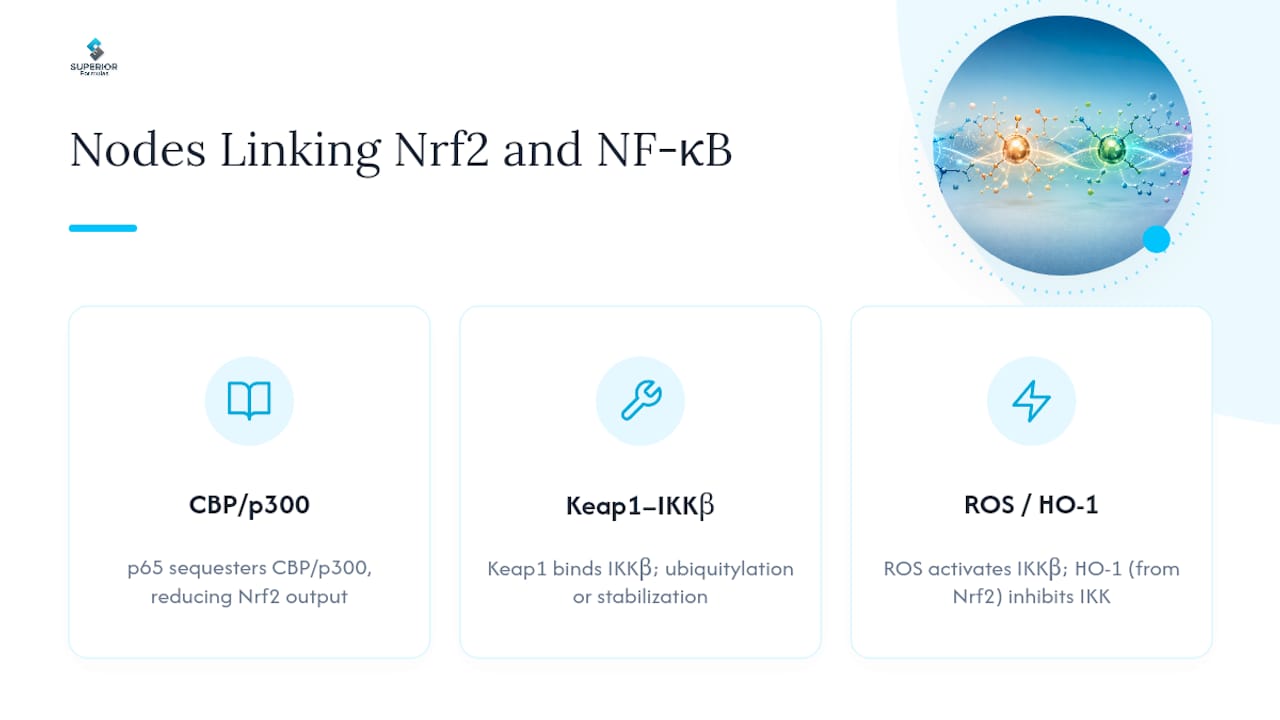
1. Competition for CBP/p300 coactivator binding. RelA/p65 and Nrf2 both require the transcriptional coactivators CBP and p300 to drive gene expression efficiently, and the two transcription factors compete for a limited nuclear pool of these proteins. When p65 is strongly activated, it can sequester CBP/p300 away from Nrf2-ARE complexes, dampening Nrf2 output independent of any change in Nrf2 protein level. Acetylation status of both factors modulates this competition further, since CBP/p300 acetylate p65 at specific lysines to enhance its DNA binding, meaning the coactivator pool is being consumed for two distinct chromatin-modifying functions simultaneously, not just used as a passive bridging protein. This molecular cross-talk between Nrf2 and NF-κB has been documented across multiple cell systems and is one of the better-supported inhibitory mechanisms in the literature.
2. Keap1–IKKβ physical interaction. Keap1 does more than regulate Nrf2. It also binds IKKβ, and this interaction has been proposed to work through an ETGE-like motif on IKKβ that mimics the docking sequence Keap1 normally uses on Nrf2 itself. The functional consequence is debated. Some evidence suggests Keap1 promotes IKKβ ubiquitination and degradation, which would mean an intact Keap1 pool actually restrains NF-κB signaling independent of its Nrf2 role. Other data point toward Keap1 acting as a scaffold that stabilizes IKKβ activity under specific conditions. Either way, Keap1–IKKβ interactions represent a genuine physical node where the two systems touch directly, not just through downstream gene products, making this a priority target for proteomic follow-up.
3. ROS as a bidirectional signaling intermediate. Reactive oxygen species sit at the center of both pathways but play opposite roles depending on which system you’re asking about. Moderate ROS levels can activate IKKβ and promote NF-κB signaling, while the same ROS pool triggers Keap1 cysteine modification and Nrf2 stabilization. Once Nrf2 target genes come online, HO-1 in particular produces carbon monoxide and biliverdin, both of which have documented inhibitory effects on IKK activity and downstream cytokine transcription. This creates a negative feedback loop: NF-κB-driven inflammation generates ROS, ROS activates Nrf2, and Nrf2-driven HO-1 output then suppresses the NF-κB signal that triggered it in the first place. Whether this loop resolves quickly or gets stuck in a chronic activation state appears to depend heavily on baseline antioxidant capacity going into the stimulus.
4. GSK3β-mediated divergence in protein turnover. GSK3β phosphorylates Nrf2’s Neh6 domain to trigger β-TrCP-mediated degradation, but GSK3β activity is itself suppressed by the PI3K/Akt pathway, which is commonly activated alongside inflammatory signaling. The result is a paradox worth testing directly in any given cell system: acute inflammatory stimuli that activate Akt can simultaneously inhibit GSK3β, which would be expected to stabilize Nrf2 even while NF-κB is being actively driven. Researchers who see unexpected Nrf2 stabilization during an inflammatory time course should check GSK3β phosphorylation status before assuming a Keap1-independent artifact.
Pro Tip: When a Keap1 knockdown experiment produces an unexpected NF-κB phenotype, don’t assume it’s purely due to Nrf2 derepression. Run a parallel IKKβ stability blot, since the Keap1–IKKβ interaction can independently shift NF-κB output regardless of what’s happening at the ARE.
Context dependence ties all four nodes together. The temporal order of activation changes outcomes substantially: pre-activating Nrf2 before an inflammatory challenge tends to produce stronger NF-κB suppression than activating Nrf2 after NF-κB is already engaged, likely because the CBP/p300 pool and HO-1 induction need time to establish before they can compete effectively. Cell-type-specific expression of cofactors like small Maf proteins or NEMO isoforms also shifts the balance, and proteostasis capacity, meaning how well a cell’s autophagy and proteasome systems are functioning, determines how quickly either pathway resets to baseline after a stimulus.
What Does the Experimental Evidence Show?
Nrf2 knockout mouse models provide the clearest genetic evidence for this crosstalk, and the phenotype pattern is remarkably consistent across labs. Nrf2 deficient animals show elevated NF-κB activity and increased pro-inflammatory cytokine production compared to wild-type littermates under matched inflammatory challenge, a finding that has been replicated across multiple disease models including lipopolysaccharide-induced lung injury and chemically induced hepatotoxicity.
Nrf2-deficient mice reproducibly show heightened NF-κB pathway output and cytokine release across independent inflammatory challenge models, supporting a genuine suppressive role for Nrf2 rather than a cell-culture artifact.
Cell culture work fills in the mechanistic detail behind the whole-animal phenotype. In Nrf2-deficient cell lines, researchers have documented accelerated IκBα phosphorylation and degradation kinetics following TNFα or LPS stimulation compared to Nrf2-competent controls, consistent with loss of the HO-1-mediated brake on IKKβ activity described above. A separate line of evidence shows that active p65 can promote nuclear translocation of Keap1 itself, which would deplete cytosolic Keap1 available for its normal Nrf2-regulatory function, a mechanism that runs in the opposite direction from the more commonly cited Nrf2-suppresses-NF-κB narrative and is worth keeping in mind when interpreting any single-timepoint experiment.
Disease correlations extend this crosstalk beyond basic cell biology into contexts with direct translational stakes:
- Coronary artery disease shows documented Nrf2/NF-κB crosstalk, with Nrf2 pathway activation associated with reduced NF-κB signaling and improved vascular inflammatory markers in preclinical models, an axis now being explored for regulation by sirtuin proteins like SIRT6.
- Drug-induced organ toxicity, particularly hepatotoxicity from acetaminophen overdose, shows Nrf2-deficient models with substantially worse liver injury scores, tracking with unrestrained NF-κB-driven inflammatory infiltration.
- Neurodegeneration research links chronic microglial NF-κB activation with declining Nrf2 responsiveness in aging brain tissue, a pattern implicated in Parkinson’s and Alzheimer’s disease models where neuroinflammation is a central feature.
- Cancer biology complicates the picture further, since constitutive Nrf2 activation (often through Keap1 mutation) can paradoxically support tumor cell survival by suppressing the same inflammatory and oxidative stress signals that would otherwise trigger apoptosis, meaning the “Nrf2 good, NF-κB bad” framing breaks down entirely in oncogenic contexts.
How Can Researchers Modulate the Nrf2 and NF-κB Crosstalk?
Compound selection for crosstalk experiments needs to account for the fact that many so-called Nrf2 activators exert at least part of their effect through direct NF-κB suppression, not purely through the Keap1-ARE axis. This makes mechanism-of-action controls essential rather than optional.
Nrf2-directed compounds. Sulforaphane, an isothiocyanate derived from cruciferous vegetables, remains the most extensively characterized Keap1 cysteine modifier in the literature, producing robust ARE-luciferase activation at low micromolar concentrations in most cell lines. Several polyphenols, including curcumin and resveratrol, show dual activity, activating Nrf2 through mild electrophilic stress while also directly inhibiting IKKβ phosphorylation, which means any observed NF-κB suppression with these compounds cannot automatically be attributed to Nrf2-mediated HO-1 induction without a knockdown control. The broader literature on natural compounds modulating both Nrf2 and NF-κB pathways documents this dual mechanism across a wide range of polyphenol classes, reinforcing why single-pathway conclusions from compound screens deserve skepticism.
NF-κB-directed compounds. IKKβ inhibitors like BMS-345541 offer more pathway-specific NF-κB suppression than polyphenol treatment, making them useful as a mechanistic anchor when trying to isolate Nrf2-independent effects. Proteasome inhibitors such as bortezomib block IκBα degradation and thereby suppress NF-κB nuclear translocation, but they simultaneously block Keap1-mediated Nrf2 degradation, which stabilizes Nrf2 as a direct side effect of the treatment rather than through genuine pathway crosstalk. This confound trips up more compound screens than it should. Broad, sustained NF-κB inhibition also carries real biological risk given that NF-κB signaling has documented pro- and anti-inflammatory roles depending on cellular context, so chronic blockade in animal models can produce unexpected immunosuppressive or tissue-repair deficits that complicate interpretation of the primary endpoint.
Recommended experimental design points for anyone building a crosstalk protocol:
- Stagger co-treatment timing (pre-treatment versus simultaneous versus post-challenge) since the temporal order changes outcomes substantially, as noted above.
- Run full dose-response curves for any Nrf2 activator being tested for NF-κB effects, since many compounds show biphasic activity where low doses activate Nrf2 cleanly but higher doses introduce direct cytotoxic or off-target IKK effects.
- Pair antioxidant gene readouts (HO-1, NQO1) with cytokine readouts (TNFα, IL-1β) in the same experiment rather than relying on one pathway’s output as a proxy for the other.
- Include an HO-1 inhibitor arm (such as tin protoporphyrin) alongside any Nrf2 activator when trying to establish whether observed NF-κB suppression is genuinely HO-1-dependent.
Pro Tip: If your Nrf2 activator suppresses NF-κB output but that suppression disappears when you co-administer an HO-1 inhibitor, you’ve isolated the mechanism to the HO-1/carbon monoxide axis rather than a direct Keap1–IKKβ or CBP/p300 effect. That distinction changes which downstream biomarker you should track in vivo.
What Are the Open Questions in Nrf2 and NF-κB Crosstalk Research?
Several mechanistic questions remain genuinely unresolved, and they matter for anyone designing the next round of experiments in this space.
The composition and dynamics of the Keap1–IKKβ complex under live conditions is still poorly characterized. Most evidence comes from co-immunoprecipitation snapshots rather than real-time tracking, leaving open whether the interaction is constitutive or stimulus-triggered. Tissue-specific wiring is another gap: nearly all of the mechanistic work described above comes from a handful of cell lines and liver or lung tissue in rodent models, and whether the same node hierarchy holds in neurons, cardiomyocytes, or immune cell subsets specifically is largely untested.
Three experimental strategies would meaningfully advance the field:
- Proteomic mapping of the Keap1 interactome under basal and inflammatory-stimulated conditions, to determine whether IKKβ binding is constitutive or induced and how it compares across cell types.
- Dual live-cell reporter systems pairing an ARE-luciferase or fluorescent Nrf2 sensor with an NF-κB reporter in the same cells, enabling real-time tracking of both pathways’ temporal relationship rather than inferring it from separate endpoint experiments.
- Conditional, tissue-specific knockout models for Keap1 or IKKβ, paired with single-cell RNA sequencing, to resolve whether the crosstalk hierarchy identified in liver and macrophage studies generalizes to neural or cardiovascular tissue.
The most common interpretive pitfall is relying on a single marker, usually HO-1 or phospho-p65, as a stand-in for whole-pathway activity, when both systems have multiple parallel readouts that can diverge from each other over time.
What an Experimenter’s Checklist for This Crosstalk Should Include
Three habits separate a defensible crosstalk finding from an artifact. First, use orthogonal readouts: never conclude Nrf2 activation from a reporter assay alone without confirming nuclear translocation and downstream target gene induction independently. Second, sample across time, not at a single endpoint, since the CBP/p300 competition and HO-1 feedback loop described above unfold over hours, and a snapshot at one timepoint can catch either pathway mid-transition. Third, match your controls: vehicle, genetic knockdown, and pathway-specific inhibitor arms all need to run in parallel, not sequentially across different experiments.
Moving from cell culture to animal models deserves particular caution here. A mechanism confirmed in one cell line, even with clean orthogonal readouts, does not automatically generalize to whole-organism inflammation, where systemic cytokine exposure, immune cell trafficking, and tissue-specific cofactor expression introduce variables no monoculture system can capture. Treat cellular crosstalk data as hypothesis-generating for the in vivo work, not as a stand-in for it.
— cristopher
Where Standardized Nrf2 Support Fits Into Translational Research
Researchers evaluating Nrf2-directed compounds for translational or adjunct use often run into the same practical obstacle: sourcing consistency. A polyphenol blend with a batch-to-batch shift in sulforaphane or fisetin content will quietly undermine a dose-response experiment before the biology even gets tested.

Superiorformulas builds its Nrf2-focused formulations, including Formula 1’s Longevity Daily Antioxidant Blend, on physician-formulated recipes manufactured in GMP-certified facilities with third-party testing for purity and potency. That level of standardization is exactly what dose-response and vehicle-matched control work depends on. For anyone designing a translational protocol around Nrf2 activation or reviewing the broader science behind these formulations, consistent sourcing removes one major confound before the experiment starts. Clinicians recommending an evidence-informed antioxidant option for patients, meanwhile, can review the full formulation lineup and match ingredient specifications against their own protocol requirements before making a recommendation.
Sources
- Dissecting molecular cross-talk between Nrf2 and NF-κB response pathways
- The crosstalk between Nrf2 and NF-κB pathways in coronary artery disease: Can it be regulated by SIRT6?
- NF-κB signaling in inflammation | Signal Transduction and Targeted Therapy
- Modulation of Nrf2 and NF-κB Signaling Pathways by Naturally Occurring Compounds
FAQ
Does NF-κB Increase Inflammation?
Yes, canonical NF-κB activation drives transcription of pro-inflammatory cytokines like TNFα and IL-1β, but its role is context-dependent. NF-κB also supports tissue repair and immune cell survival, so blunt, sustained inhibition can produce unintended effects beyond reducing inflammation.
Does Nrf2 Reduce Inflammation?
Nrf2 activation generally reduces NF-κB-driven inflammation by inducing antioxidant genes like HO-1 and by competing directly with p65 for CBP/p300 coactivators. The magnitude of that suppression depends on cell type, timing, and baseline oxidative stress levels.
Is NF-κB Good or Bad?
Neither label fits cleanly. NF-κB is essential for acute immune defense and tissue repair, but chronic, unresolved activation is strongly associated with neurodegenerative and cardiovascular disease, making duration and context more important than the pathway’s mere presence.
What Foods Trigger Nrf2 Activation?
Cruciferous vegetables like broccoli and Brussels sprouts supply sulforaphane, one of the best-characterized natural Keap1 modifiers, while foods rich in polyphenols such as curcumin and resveratrol also show documented Nrf2-activating effects. Researchers interested in a standardized, dose-consistent source rather than variable dietary intake can review Nrf2-targeted formulations built around these same compound classes.
What Is the Main Difference Between Nrf2 and NF-κB Function?
Nrf2 primarily drives cytoprotective, antioxidant gene expression to help cells manage oxidative stress, while NF-κB primarily drives inflammatory and immune-response gene expression in reaction to pathogens or tissue damage. The two systems intersect at multiple points, including Keap1–IKKβ binding and competition for shared coactivators, rather than operating in isolation.


